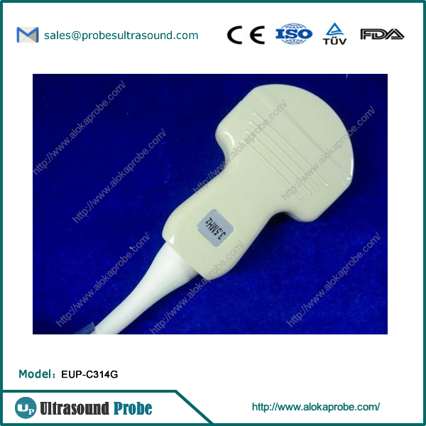
File:2-2 EUP-C314G.jpg
Ultrasonogarphy is increasingly being used in clinical diagnostic settings and quickly replacing other radiologic methodologies. X rays have deleterious effects on tissues that are being exposed to them. They are also ineffective when keen imaging of fluid and tissues are needed. So ultrasound offers the advantage of being a safe procedure while providing detailed images of tissues that can aid in the diagnostic process. Commonly, the picture conjured up when ultrasound technician is mentioned is one that performs ultrasound testing on fetuses, which is just the tip of the iceberg.hdmi extender Due to its non-invasive nature, ultrasonography has a wide range of uses that go beyond monitoring the growth of a fetus. Even the smallest facets of anatomy can be analyzed to create an image both for diagnosis and understanding complex mechanisms of function.
Diagnostic medical sonographers may dedicate themselves to one or more of specialized disciplines within ultrasound technician career path. The specializations includeAbdominal sonography, which deals with studies of the spleen, liver, pancreas and gall bladderObstetric and gynecologic sonography, dealing with the female reproductive systemCardiacsonography, involving the study of muscle tissue in the heartVascular sonography, involving analysis of blood flow in veins and arteriesNeurosonography, that deals with studying brain tissueBreast sonography, that is focused on analyzing breast tissue andOphthalmologic sonography that studies eyesAbdominal Sonography
Abdominal ultrasound technicians study the conditions that require the imaging of the spleen, bile duct, gall bladder, kidneys and pancreas. Note that lungs cannot be analyzed with sonography as sound waves produce poor echoing patterns with gases and bones.Obstetric and Gynecologic Sonography
Ultrasound technicians that specialize in obstetrics and gynecologists create and analyze images of the fetus and the female reproductive system. The ultrasound tech performs calculations of head circumference, length of tibia among others and prepares a report for the physician who will then discuss the details and prognoses with the patient. This is helpful in finding birth defects and abnormal growths in pregnant women. Conditions such as the thickness of the lining of the uterus along with monitoring ovulation also fall under the purview of what an obstetric sonographer will do.Cardiac Sonography
Performing echocardiograms which is the study of heart tissue and functioning is the primary role of the cardiac ultrasound technician. The blood flow through and around the heart are used to diagnose heart conditions. Real-time imaging using Doppler ultrasound is used to study the real time functioning of the heart. An in-depth analysis of the heart valves and muscle tissue are vital in diagnosing cardiac conditions.Vascular Sonography
Vascular ultrasound technicians are valuable in the examination of complex veins and arteries resulting in the diagnosis of abnormalities or diseases such aneurisms and peripheral arterial disease. Conditions of the veins of the upper and lower extremities that cause pulmonary embolisms are also an integral part of vascular sonography. Detecting blockages or coagulations through vascular sonography is a common use for patients with clotting issues.Neurosonography
Neurosonographers study diseases of the central nervous system. Nervous system blood vessels are examined in order to diagnose stroke and brain aneurysms. Neurosonography is particularly useful in diagnosing neurological disorders in premature infants. Conditions that cause stroke such as sickle cell anemia are detected by Neurosonography. The transducers and frequencies employed are different for neursonographers than abdominal ultrasound techs for instance.Breast Sonography
Breast ultrasound technicians examine tissues of the breast to detect abnormalities and disease. These tests are referred to as mammograms, but these tests are limited in that they can only detect inconsistencies in identifying whether the inconsistency is solid or fluid. Beyond this point, a needle biopsy might have to be performed to determine if the abnormality is benign or malignant. Ultrasonography is used to guide the needle to its target tissue. Once retrieved the cells are studied under the microscope to detect malignancy if present.Opthalmologic Sonography
Opthalmologic ultrasound technicians are used to study eyes. Prosthetic lenses are inserted in the eye with the guidance of Ultrasonography. This allows for accurate measurements. Since the eye and the region surrounding the eyes are mostly fluid and muscle tissue, ultrasonography is a valuable instrument in detecting separated retinas, blood supply interruptions and other diseases of tissues surrounding the eye. Here the transducers are much smaller and specially designed for study of the eye.
File history
Click on a date/time to view the file as it appeared at that time.
| Date/Time | Thumbnail | Dimensions | User | Comment | |
|---|---|---|---|---|---|
| current | 20:10, 17 April 2016 |  | 600 × 600 (176 KB) | Dfssgfwsdre (Talk | contribs) | Ultrasonogarphy is increasingly being used in clinical diagnostic settings and quickly replacing other radiologic methodologies. X rays have deleterious effects on tissues that are being exposed to them. They are also ineffective when keen imaging... |
- You cannot overwrite this file.
File usage
There are no pages that link to this file.
